|

| Home | Crystal | jmol | jPOWD | Chem | X Ray | Dana | Strunz | Properties | A to Z | Images | Share | News | Help | About |
Images from American MineralogistClick on thumbnails for a larger image. Please be patient since some of these pages of thumbnail images take some time to load. These mineral photos are copyrighted by their owners and may not be used without permission. Pages: [1] [2] [3] [4] [5] [6] [7] [8] [9] [10] [11] [12] [13] [14] [15] [16] [17] |
Okayamalite Image | |||
|
|
|
||
Okayamalite Image | |||
|
|
|
||
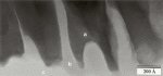
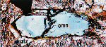
Ominelite Image | |||
|
|
|
||
Ominelite Image | |||
|
|
|
||
Orickite Image | |||
|
|
|
||
Orickite Image | |||
|
|
|
||
Paulkellerite Image | |||
|
|
|
||
Paulkellerite Image | |||
|
|
|
||
Paulmooreite Image | |||
|
|
|
||
Paulmooreite Image | |||
|
|
|
||
Phosphovanadylite Image | |||
|
|
|
||
Phosphovanadylite Image | |||
|
|
|
||
Piergorite-(Ce) Image | |||
|
|
|
||
Piergorite-(Ce) Image | |||
|
|
|
||
Polhemusite Image | |||
|
|
|
||
Polhemusite Image | |||
|
|
|
||
Poppiite Image | |||
|
|
|
||
Poppiite Image | |||
|
|
|
||
Protoanthophyllite Image | |||
|
|
|
||
Protoanthophyllite Image | |||
|
|
|
||
Small.jpg)
Pages: [1] [2] [3] [4] [5] [6] [7] [8] [9] [10] [11] [12] [13] [14] [15] [16] [17] |