American Mineralogist's Image Gallery

American Mineralogist's Image Gallery

Search Webmineral :
   
Home Crystal jmol jPOWD Chem X Ray Dana Strunz Properties A to Z Images Share
      News Help About

Images from American Mineralogist

Click on thumbnails for a larger image. Please be patient since some of these pages of thumbnail images take some time to load. These mineral photos are copyrighted by their owners and may not be used without permission.

Pages: [1] [2] [3] [4] [5] [6] [7] [8] [9] [10] [11] [12] [13] [14] [15] [16] [17]
American Mineralogist's Image Gallery With 326 Images

Schreyerite Image

Click Here for Larger Image in New Browser Window Images:
Click Here for Larger Image

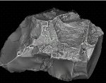
Schreyerite: V+++2Ti3O9

Comments: SEM image of schreyerite crystal studied by single-crystal X-ray diffraction. AmMin, v91, p197.
Location: Metamorphic rocks of the Sludyanka complex at the southern shore of Lake Baikal, Russia. Scale: Picture width 0.14 mm.
© American Mineralogist

Serrabrancaite Image

Click Here for Larger Image in New Browser Window Images:
Click Here for Larger Image

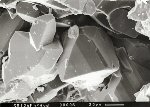
Serrabrancaite: MnPO4·(H2O)

Comments: SEM micrograph of dark brown to dark greenish black isometric crystals of serrabrancaite up to 0.3 mm size. (AmMin, 85:848).
Location: Alto Serra Branca pegmatite near Pedra Lavrada, Paraiba, Brazil. Scale: See Image.
© American Mineralogist

Shirozulite Image

Click Here for Larger Image in New Browser Window Images:
Click Here for Larger Image

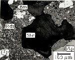
Shirozulite: KMn3(AlSi3)O10(OH,F)2

Comments: Back-scattered-electron image of shirozulite. Shr = shirozulite; Tp = tephroite; Ap = apatite; Rds = rhodochrosite; Bm = bementite. (AmMin, 89:233).
Location: Tauchi mine, Aichi Prefecture, Japan. Scale: See Image.
© American Mineralogist

Silicon Image

Click Here for Larger Image in New Browser Window Images:
Click Here for Larger Image

Silicon: Si  
Moissanite: SiC

Comments: Backscattered electron image of metallic inclusion in moissanite (SiC). The gray shading in the BSE mage consists mainly of pure silicon (Si metal). The brighter areas are composed of Fe-silicide. (AmMin, 88:1818).
Location: Found in a cobble at a beach along the Turkish coast of the Mediterranean Sea, around 150 km NW from Izmir, and is most likely derived from Tertiary volcanic rocks outcropping in the area. Scale: Scalebar 200 µ-m.
© American Mineralogist

Simmonsite Image

Click Here for Larger Image in New Browser Window Images:
Click Here for Larger Image

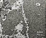
Simmonsite: Na2LiAlF6

Comments: Backscattered electron images of primary assemblage of cryolite-cryolithionite (cl) simmonsite (sm) and trace elpasolite (ep). (AmMin, v 84, p 770).
Location: Zapot amazonite-topaz-zinnwaldite pegmatite located in the Gillis Range, Mineral Co., Nevada, USA. Scale: See Image.
© American Mineralogist

Sorosite Image

Click Here for Larger Image in New Browser Window Images:
Click Here for Larger Image

Sorosite: Cu(Sn,Sb)  
Tin: Sn  
Stistaite: SnSb

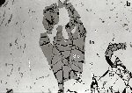
Comments: Back-scattered electron image of large sorosite crystal (So=partly skeletal) adjacent to stistaite (St). Tn=native tin. Note abundant microcrystals of sorosite isolated within native tin. (AmMin 83:903).
Location: Baimka gold-platinum group mineral placer deposit, Chukotka, Russian Far East. Scale: Image Size 1.5 cm.
© American Mineralogist

Spriggite Image

Click Here for Larger Image in New Browser Window Images:
Click Here for Larger Image

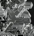
Spriggite: Pb3[(UO2)6O8(OH)2](H2O)x; x ~ 3

Comments: Secondary electron microscope image of spriggite crystals and twins. (AmMin, 89:341).
Location: Number 2 workings, Radium Ridge, Northern Flinders Ranges, South Australia. Scale: See Image.
© American Mineralogist

Srilankite Image

Click Here for Larger Image in New Browser Window Images:
Click Here for Larger Image

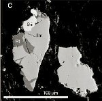
Srilankite: (Ti,Zr)O2

Comments: Backscatter electron image of srilankite (Sri) - rutile (Rt) - ilmenite (Ilm) composite grain (AmMin, v 89, p 761).
Location: Atlantis II Fracture Zone, Southwest Indian Ridge, Southern Indian Ocean. Scale: See Image.
© American Mineralogist

Stornesite-(Y) Image

Click Here for Larger Image in New Browser Window Images:
Click Here for Larger Image

Stornesite-(Y): (Y, Ca)[ ]2Na6(Ca,Na)8(Mg,Fe)43(PO4)36

Comments: BSE image of stornesite-(Y) (Sto) partially surrounding a spherical quartz(Qz)-biotite(Bt) aggregate in apatite (Ap). Other mineral phases include xenotime (Xnt), sillmanite Sil) and corderite (Crd). AmMin, v91, p 1416.
Location: Paragneiss specimens from Johnston Fjord, Stornes Peninsula, Antarctica. Scale: See Image.
© American Mineralogist

Suessite Image

Click Here for Larger Image in New Browser Window Images:
Click Here for Larger Image

Suessite: (Fe,Ni)3Si

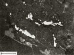
Comments: B&W image of suessite (white) as vein fillings and blebs. Silicates (olivine, pigeonite) are gray; carbonaceous matrix is black. Hydrous ferric oxide veins are (light gray).
Location: North Haig olivine pigeonite achondrite (ureilite). (AmMin, v67:126). Scale: Scale Bar 100 µm.
© American Mineralogist

Pages: [1] [2] [3] [4] [5] [6] [7] [8] [9] [10] [11] [12] [13] [14] [15] [16] [17]