|
")
| Home | Crystal | jmol | jPOWD | Chem | X Ray | Dana | Strunz | Properties | A to Z | Images | Share | News | Help | About |
Mineral Thumbnail Images (D)[Up] [A] [B] [C] [D Images] [E] [F] [G] [H] [I] [J] [K] [L] [M] [N] [O] [P] [Q] [R] [S] [T] [U] [V] [W] [X] [Y] [Z]Click on thumbnails for a larger image. Please be patient since some of these pages of thumbnail images take some time to load. These mineral photos are copyrighted by their owners and may not be used without permission. Pages: [1] [2] [3] [4] [5] [6] [7] [8] [9] [10] [11] [12] [13] [14] |
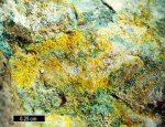
Dietzeite Image | |||
|
|
|
||
Dietzeite Image | |||
|
|
|
||
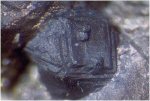
Digenite Image | |||
|
|
|
||
Digenite Image | |||
|
|
|
||
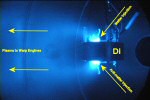
Dilithium Image | |||
|
|
|
||
Dilithium Image | |||
|
|
|
||
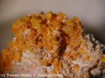
Dimorphite Image | |||
|
|
|
||
Dimorphite Image | |||
|
|
|
||
Diopside Image | |||
|
|
|
||
Diopside Image | |||
|
|
|
||
Diopside Image | |||
|
|
|
||
Diopside Image | |||
|
|
|
||
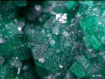
Dioptase Image | |||
|
|
|
||
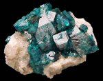
Dioptase Image | |||
|
|
|
||
Dioptase Image | |||
|
|
|
||
Dioptase Image | |||
|
|
|
||
Dioptase Image | |||
|
|
|
||
Dioptase Image | |||
|
|
|
||
Dioptase Image | |||
|
|
|
||
Dioptase Image | |||
|
|
|
||
Pages: [1] [2] [3] [4] [5] [6] [7] [8] [9] [10] [11] [12] [13] [14] |